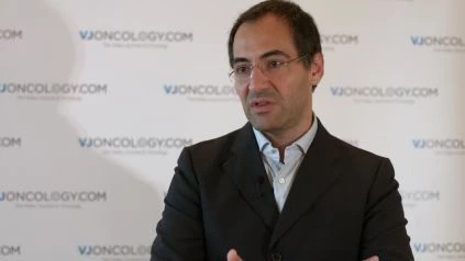
Giovanni Pellacani, MD of the University of Modena and Reggio Emilia, Modena, Italy, discusses his talk on imaging techniques in melanoma presented at the 2016 World Congress on Cancers of the Skin (WCCS) and the Congress of the European Association of Dermato-Oncology (EADO) in Vienna, Austria. His talk focused on new imaging techniques that enable the use of in vivo, non-invasive methods to conduct an examination of the tissue in depth, at a histological level resolution. This means that using this method, we can see the cells and the structures, similarly as when using histopathology. The tool he describes is respectively confocal microscopy. He highlights that this tool enables the exploration of the skin, and allows for a more accurate diagnosis of skin cancer, especially that of melanoma and cases where clinical and microscopic examination may prove to be difficult. Next, he discusses another tool named multiphoton microscopy. This tool is still a research tool, but proves to be promising as it can also generate images related to the metabolism of the cells. Prof. Pellacani elaborates that the cells seen here can be colored and cells that are more biologically active can be distinguished from that of the remaining cells, resulting in this tool potentially holding further positive clinical implications.
[the_ad id="32629"]

